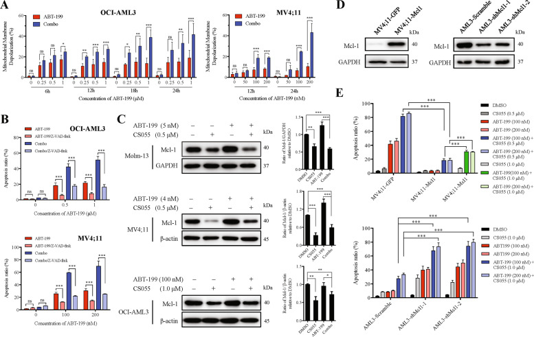
Fig. 4. CS055 enhances mitochondrion-dependent apoptosis induced by ABT-199 by downregulation of Mcl-1.
a OCI-AML3 (left) and MV4;11 cells (right) were exposed to the indicated concentrations of ABT-199 ± CS055 (1.0 μM for OCI-AML3; 0.5 μM for MV4;11) for 6–24 h, after which mitochondrial membrane potential (MMP) was measured using the JC-1 kit by flow cytometry. b OCI-AML3 (upper) and MV4;11 cells (lower) were pre-incubated with 20 μM Z-VAD-fmk for 2 h, followed by treatment with indicated concentrations of ABT-199 ± CS055 (1.0 μM for OCI-AML3; 0.5 μM for MV4;11) for additional 24 h. The percentage of apoptosis was then assessed by flow cytometry. c Western blot analysis was performed to monitor the expression of Mcl-1 in Molm-13, MV4;11, and OCI-AML3 cells treated with the indicated concentration of ABT-199 ± CS055. Blots were probed for β-actin or GAPDH as loading controls. d Mcl-1 was ectopically expressed in MV4;11 cells (left) or knocked down by shRNA in OCI-AML3 cells (right). e MV4;11 cells overexpressing Mcl-1 (upper) and OCI-AML3 cells with Mcl-1 knockdown (lower) were treated with indicated concentrations of ABT-199 ± CS055 for 48 h, after which the percentage of apoptosis was determined by flow cytometry. For panels a, b and e, values indicate mean ± SD for at least three independent experiments performed in triplicate (*P < 0.05, **P < 0.01, and ***P < 0.001, ns not significant).

