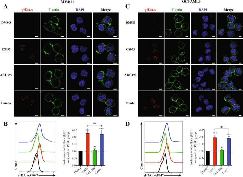
Fig. 5. CS055 induces expression and foci formation of γH2AX, an event not enhanced by ABT-199.
MV4;11 (a, b) and OCI-AML3 cells (c, d) were treated with ABT-199 (5 nM for MV4;11, 100 nM for OCI-AML3) ± CS055 (0.5 μM for MV4;11, 1.0 μM for OCI-AML3) for 18 h, after which cells were subjected to immunofluorescent staining for Ser139 phosphorylation of histone H2A.X (H2A.X, red) and confocal microscopy (a, c; phalloidin—green, DAPI—blue; scale bars: 5 μm.) or flow cytometry for monitoring H2A.X expression (b, d). Values indicate mean ± SD for at least three independent experiments performed in triplicate (***P < 0.001, ns not significant).

