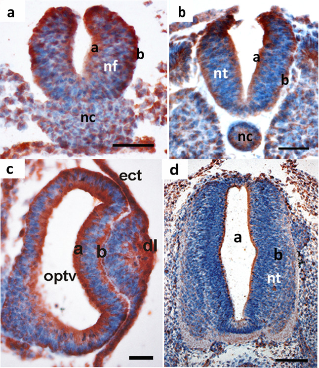
Fig. 2.

Mdg1/ERdj4 protein pattern in the developing nervous system. a HH-stage 12, coronal section. Mdg1/ERdj4 protein accumulates at the apical (a) and basal (b) sites of the neural fold (nf), which has not yet closed to form the roof plate. nc, notochord. Scale bar 50 µm. b HH-stage 13, coronal section. Mdg1/ERdj4 protein is present at comparable amounts in the basal (b) and apical (a) zone of the neural tube (nt). Scale bar 50 µm. c HH-stage 13, frontal section of the optic vesicle in the developing brain. The polarized Mdg1/ERdj4 distribution is clearly visible in the apical (a) and basal (b) zone of the brain vesicle that forms the optic vesicle (optv). ect, ectoderm; dl, developing lens. Scale bar 50 µm. d Chick embryo day 4, the apical zone of the neural tube, which consists of secretorily active ependymal cells, contains high levels of Mdg1/ERdj4 protein. Scale bar 100 µm. Scale bar 100 µm
