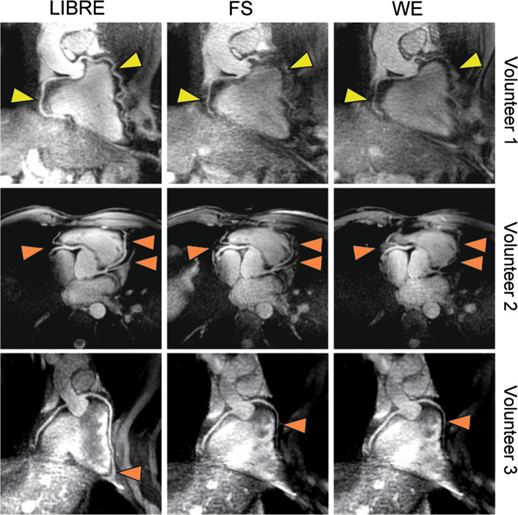
Fig. 4.
Comparison of the different fat saturation methods on radial trajectories coronary MRA at 3 T in healthy subjects. Coronary MRA images show the left and right coronary artery system depicting the RCA and the LAD in several subjects. Using the LIBRE pulse the visualization of the RCA and LAD was improved (yellow arrow), as well as fat suppression (orange arrows) compared with FS and WE. Vessel sharpness as well as imaged vessel length was significantly increased using LIBRE. Window and level are identical in images acquired in each volunteer. MRA magnetic resonance artery, RCA right coronary artery, LAD left anterior descending artery, LIBRE lipid insensitive binomial off-resonant excitation, FS fat saturation, WE water excitation.
(Reprinted with permission from Batiaansen et al. [59] Copyright © 2019 by the Authors)

