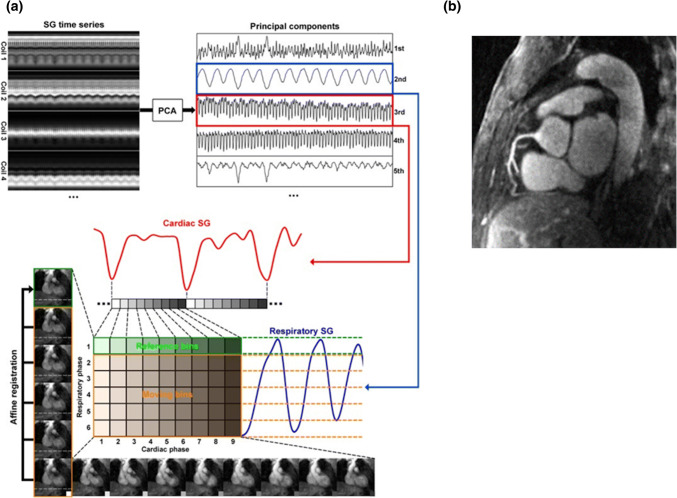
Fig. 6.
Schematics of the proposed self-gating, data binning, and respiratory motion correction framework. a First, the cardiac and respiratory motion components are identified from the PCA of the multichannel self-gating profile time series. Then, the imaging data are mapped to different cardiac and respiratory bins based on its cardiac and respiratory phase derived from the motion signals. Next, with one common respiratory phase selected as reference (in this example, respiratory phase 1 for cardiac phases 1–9), all other bins (respiratory phases 2–6, cardiac phases 1–9) are registered to the corresponding reference bin of the same cardiac phase using an affine transform model. The k-space trajectory and data are then modified accordingly for respiratory motion correction. The six images on the left show the six respiratory phases in cardiac phase 1. The horizontal dashed lines help visualize the SI motion of the heart due to respiration. The nine images on the bottom show the nine cardiac phases in respiratory phase 6. The contraction of the left ventricle can be clearly seen. b A representative case with excellent depiction of the right coronary artery. PCA principal component analysis.
Reprinted with permission from Pang et al. [17] Copyright © 2014 by the Wiley Periodicals, Inc.

