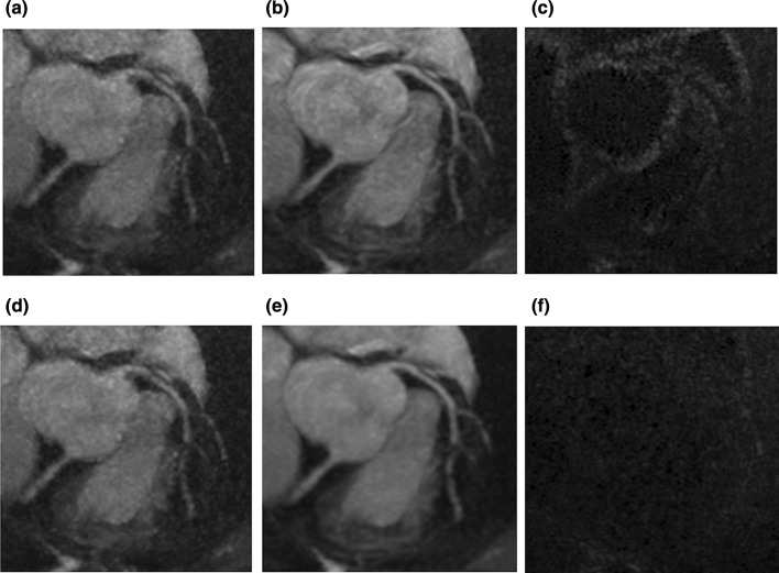
Fig. 7.
Comparison of subtracted noises by conventional denoising and DLR on the same coronary MRA data set. a, d Same original coronary MRA images acquired with parallel imaging acceleration technique. b Coronary MRA image after conventional denoising. e Coronary MRA image after DLR. c The subtracted noise between a and b. f The subtracted noise between d and e. All the panels a–f are in the same WL and WW. Subtraction images show that conventional denoising removes the edges from the original coronary MRA image (c), while DLR works on noise and retains the edge information (f). Consequently, the signal-to-noise ratio is higher in DLR processed image (e) than the conventional denoised image (b). The original image was acquired with a spoiled gradient echo sequence with ECG-gating, diaphragm navigator gating, and fat suppression with spectral attenuated inversion recovery. The MRI acquisition parameters were FOV = 380 × 380 mm, matrix = 392 × 384, slice thickness = 1.0 mm, slice number = 152, acceleration factor = 2.0 × 2.0, TR = 5.3 ms, TE = 2.0 ms, flip angle = 12 degree, bandwidth = 279 Hz/pix, acquisition voxel size = 1.0 × 1.0 × 1.0 mm, reconstructed voxel size = 0.5 × 0.5 × 0.5 mm, navigator gating window = 4 mm. For the conventional filtering, GA01 filter was used. For the DLR technique, see references [116, 140]. DLR deep learning reconstruction, MRA magnetic resonance angiography, WL window level, WW window width, GA gain algorithm

