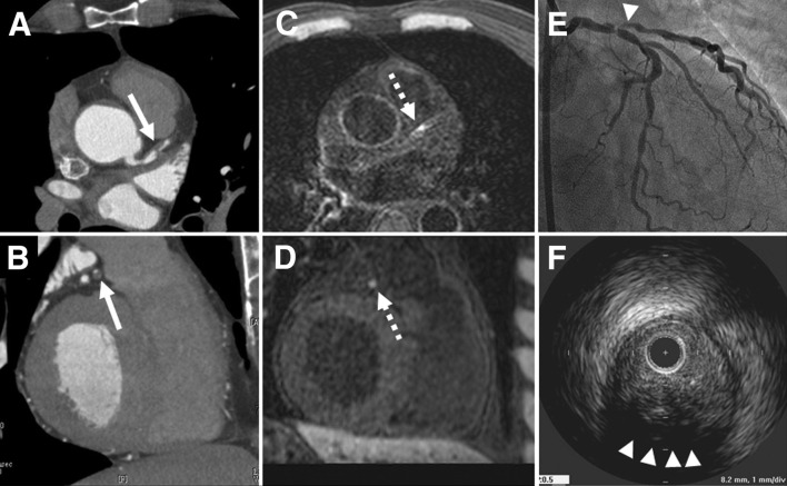
Fig. 9.
Representative case of HIP in the proximal LAD is presented. Coronary CTA (a horizontal, b sagittal) demonstrates the low-density positive remodeling plaque (− 32 Hounsfield units, remodeling index: 1.27) (arrow) with severe coronary stenosis in the proximal LAD. On the corresponding CMR (c horizontal, d sagittal), this low-density plaque was visualized as a “hyperintense spot” (dashed arrow). On the coronary angiography, severe coronary stenosis was observed (e) (arrowhead), and on IVUS examination (f), positive remodeling plaque (remodeling index: 1.29) with ultrasound attenuation (arrowheads) was observed in the proximal LAD, corresponding to the plaque observed by both coronary CTA and CMR. HIP hyperintense plaque, LAD left anterior descending artery, CTA computer tomography angiography, CMR cardiac magnetic resonance, IVUS intravascular ultrasound.
(Reprinted with permission from Kawasaki et al. [121] Copyright © 2009 by the American College of Cardiology Foundation)

