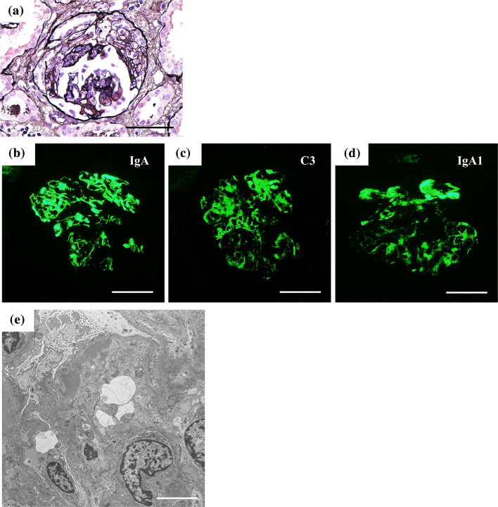
Fig. 3.
Kidney biopsy findings of the follow-up biopsy. Light microscopy uncovered fibrocellular crescent formation (a). Periodic acid-Schiff stain; Intense signals were detected for IgA (b), C3 (c), and IgA1 (d) in the mesangial areas and along the capillary walls. Scale bars = 50 μm. e Foot process effacement, microvilli degenerations, and persistent electron-dense deposits in the subendothelial and mesangial areas were observed on electron microscopy (Scale bar = 20 μm)

