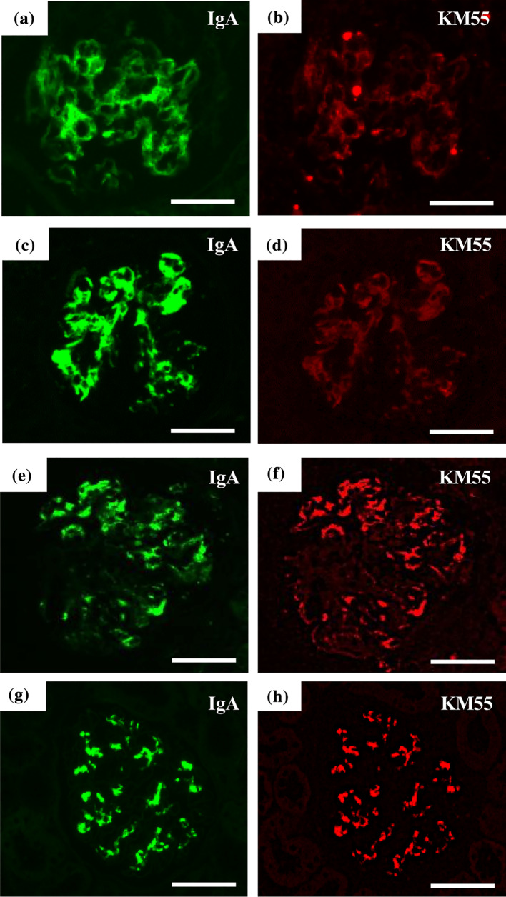
Fig. 4.
Immunofluorescence study of IgA and KM55. Immunofluorescence study of IgA and KM55 were performed simultaneously in the first and follow-up biopsy specimens of the present case (a–d) and in glomeruli of two patients with IgA nephropathy (e–h). Some amount of IgA (a) was co-localized with KM55 (b) in the glomeruli of the first biopsy specimen. The follow-up biopsy specimen showed similar findings (c, d). IgA and KM55 were co-localized in two patients with IgA nephropathy (e–h). Signals for KM55 in both the first and the follow-up biopsy specimens of the present case were weaker than those in the glomeruli of patients with IgA nephropathy (b, d, f, h). Scale bars = 50 μm

