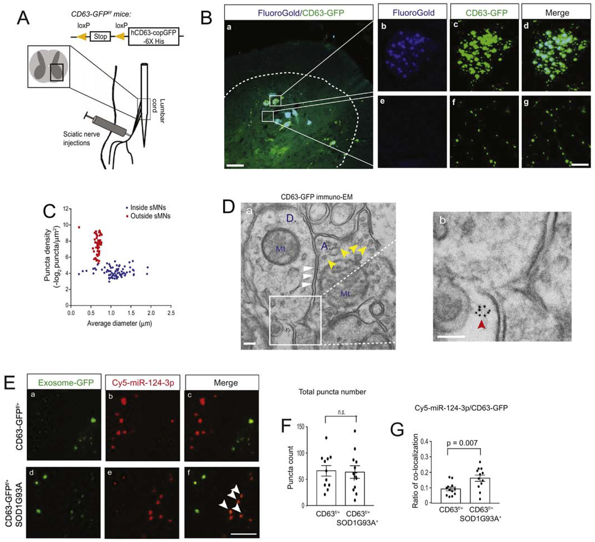
Figure 3. Increased association between miR-124–3p and motor neuron-derived exosomes in lumbar cord of SOD1G93A mice.

(A) Diagram of peripheral sciatic nerve injections of AAV9-CaMKII-Cre/FG or AAV9-CaMKII-Cre/FG/Cy5-miR-124–3p to CD63-GFPf/+ and CD63-GFPf/+SOD1G93A mice. (B) Representative images of AAV9-CaMKII-Cre-induced CD63-GFP expression and FG labeling of motor neurons in the lumbar cord of CD63-GFPf/+ mice; the dashed line shows the grey matter of lumbar cord; Scale bar: 50μm (a) and 10μm (b-g); magnified images to show intracellular and extracellular CD63-GFP labeling; (C) Quantification of the density and size of CD63-GFP+ puncta in AAV9-CaMKII-Cre and FG injected lumbar cord. Extracellular density was calculated by the total number of extracellular puncta divided by the total extracellular area of CD63-GFP+ puncta. Intracellular density was calculated by the total number of intracellular puncta divided by the individual FG+ spinal motor neurons area. Both extra- and intracellular density was –log2 converted; N = 30–40 images/4 mice/group; (D) Representative immuno-EM of CD63-GFP in the spinal cord of CaMKII-CreER+CD63f/+ mice. Immunogold labeling of CD63-GFP was performed on the CaMKII-CreER+CD63f/+ mouse spinal cord. Red arrow: extracellularly localized CD63 puncta; white arrows: post-synaptic density area; yellow arrows: synaptic vesicles; Mt: mitochondria; A: axonal terminal; D: dendritic spine; Scale bar: 100 nm. (E) Representative confocal images of CD63-GFP+ exosomes and Cy5-miR-124–3p following the injection of a mixture of FG/AAV9-CaMKII-Cre/Cy5-miR-124–3p into the sciatic nerves of CD63-GFPf/+ and CD63-GFPf/+SOD1G93A mice at P60. White arrows: co-localized CD63-GFP+ exosomes and Cy5-miR-124–3p; Scale bar: 5 μm; Quantification of the total number of extracellular CD63-GFP+ exosomes (F) and the ratio (G) of co-localized Cy5-miR-124–3p puncta with CD63-GFP+ exosomes; N = 12 images/4 mice/group, p value was determined using the two-tailed Student’s t-test.
