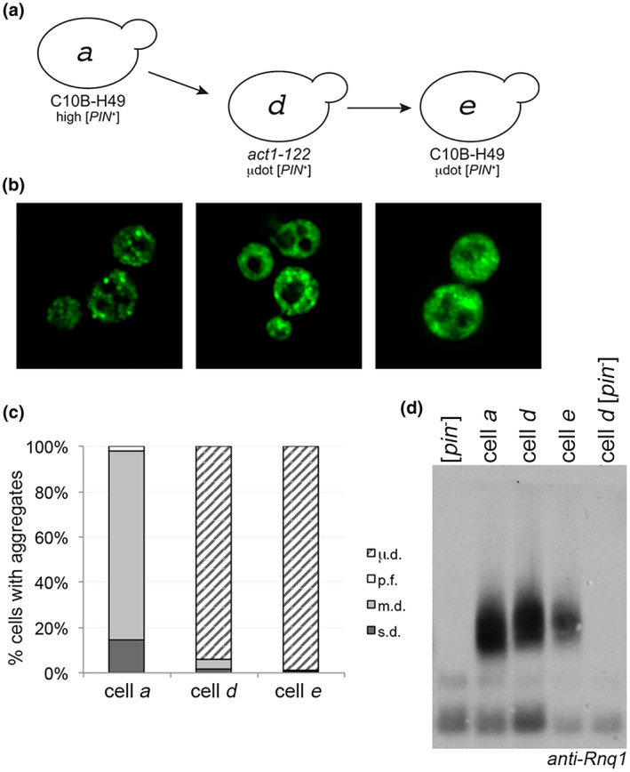
FIGURE 3.

Microdot phenotype does not revert to multiple dots when introduced into the C10B‐H49 background. (a) µdot [PIN +] in the act1‐122 strain (cell d) was cytoduced into the C10B‐H49 background (cell e). (b) Fluorescent images of cells a, d, and e. (c) Similar to Figures 1b and 2c, each bar represents the mean of at least four independent cultures and a minimum of 300 cells per culture. (d) Western blot of lysates of the indicated strains, including wildtype BY4741 [pin −] in the first lane, run on SDD‐AGE gels. Anti‐Rnq1 antibody was used to detect Rnq1 oligomers [Colour figure can be viewed at wileyonlinelibrary.com]
