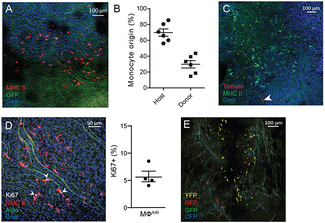
Figure 4.
Aorta intima resident macrophages (MacAIR) maintain independent of circulating progenitors and proliferate within the tissue.
(A) Parabiosis was performed to surgically link the microvasculature of CX3CR1gfp/+ reporter mice with C57BL/6 for 2 weeks, then sacrificed and assessed for transfer of cells between animals. Whole-mount immunostaining of MHC II (red) shows no cross-over of GFP+ cells between parabionts. (B) Blood analysis of 2-week parabiotic pairs shows donor/host ratios for monocytes. (C) Parabiosis was performed surgically linking the microvasculature of (cre-negative) Rosa26-mTmG reporter mice (all cells Tomato+) with C57BL/6 for 5 months, then sacrificed and assessed for cellular turn over. Whole mount confocal imaging of MHCII (green) identified resident macrophages and rare Tomato+ (red) cells were observed. (D) Aorta from C57BL/6 mice were stained for Ki67 (white) and MHC II (red) expression, imaged by confocal microscopy, and counted for the frequency of Ki67+ cells within the MacAIR population in the intima. (E) CX3CR1creER Rosa26-lsl-Confetti mice were pulsed with tamoxifen to induce recombination of stochastic fluorescent reporter alleles. Mice were left on chow diet for 4 weeks, then assessed by confocal imaging for clustering of cells of similar color to represent cellular proliferation within the tissue. Data are representative of (A) n=9 from three independent experiments, (B) representative of n=6 pairs from three independent experiments, (C) n=5 mice from two independent experiments, (D) n=4 mice across two independent experiments, and (E) n=3 from independent experiments. Error bars on graphs are presented as SEM.

