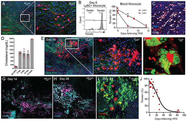
Figure 6.
Fate-mapping aorta intima resident macrophages in the progression of atherosclerosis.
(A) CX3CR1creER Rosa26-lsl-Tomato Ldlr−/− mice were treated with tamoxifen by oral gavage three times over a week, then sacrificed and imaged by en face whole mount confocal microscopy. (B) Blood monocyte labeling efficiency following tamoxifen treatment of CX3CR1creER Rosa26-lsl-Tomato Ldlr−/− mice, showing Tomato expression as assessed by flow cytometry for classical monocytes (CD115+ CD11b+ Ly6G− Ly6C+) at day 0 (left panel). Labeling kinetics for classical and non-classical monocytes in the blood tracked for 14 days after tamoxifen treatment (right panel). (C) Following tamoxifen treatment, CX3CR1creER Rosa26-lsl-tomato Ldlr−/− mice were rested on chow diet for three weeks, then assayed by whole mount en face confocal microscopy for Tomato (red) labeling of MacAIR co-stained with CD45 (white). (D) Serum cholesterol levels of CX3CR1creER Rosa26-lsl-tomato Ldlr−/− mice following labeling regiment and started on HFD feeding.(E-I) Following tamoxifen treatment, CX3CR1creER Rosa26-lsl-tomato Ldlr−/− mice were rested on chow diet for three weeks, then fed HFD for 10-days (E-F), 14 days (G), 28 days (H), and 84 days (I). MacAIR were tracked based on expression of Tomato and recruited cells were stained by antibody labeling, as indicated on the micrographs. (J) Following MacAIR labeling, the frequency of Tomato+ cells was calculated across the course of disease, from 0-84 days of HFD feeding. Data are derived from (A) n=9 from three independent experiments, (B) n=6 from two independent experiments, (C) n=6 from two independent experiments, (D) n=13 from a single experiment, (E-J) n=26 across all time points from three independent experiments. Error bars are presented as SEM.

