Fig. 3.
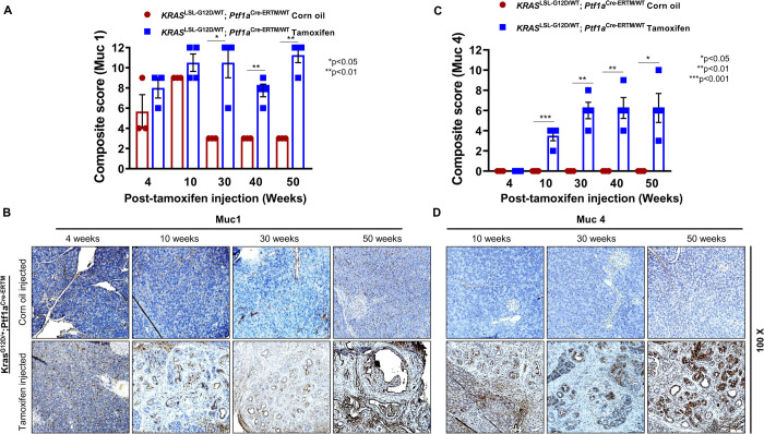
Expression of transmembrane mucins Muc1 and Muc4 in the iKC mouse model. IHC studies were performed to analyze Muc1 and Muc4 protein expression during the progression of PC in the iKC model. (A) Composite IHC scores for Muc1 show a significant increase from 10 to 50 weeks after tamoxifen injection. (B) Muc1 was expressed in iKC mice treated with corn oil or tamoxifen but was higher in tamoxifen-treated mice compared to the control mice at each time point. (C) Composite IHC scores for Muc4 show a significant increase from 10 to 50 weeks after tamoxifen injection, with no expression in corn-oil-treated mice. (D) Muc4 expression was not detected in pancreatic tissues obtained from corn-oil-treated iKC mice (upper row) or iKC mice treated with tamoxifen for only 4 weeks. Muc4 expression was detected in iKC pancreatic tissues at 10, 20, 30 and 50 weeks after tamoxifen treatment (bottom row). Muc4 expression significantly increased with cancer progression.

