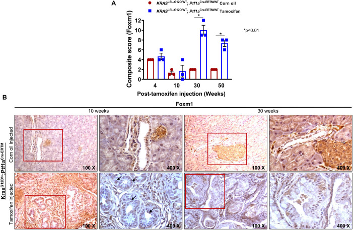
Fig. 6.
Expression of FoxM1 in iKC mice after 10 and 30 weeks of tamoxifen treatment. IHC analysis using FoxM1 antibody on pancreatic tissues isolated from corn-oil- and tamoxifen-treated iKC mice. (A) Bar graph showing quantification of immunohistochemical evaluation of FOXM1 expression in pancreas excised from corn-oil- and tamoxifen-treated mice. (B) The expression of FoxM1 in the nucleus was low in the corn-oil group (basal level), but it increased significantly upon tamoxifen injection.

