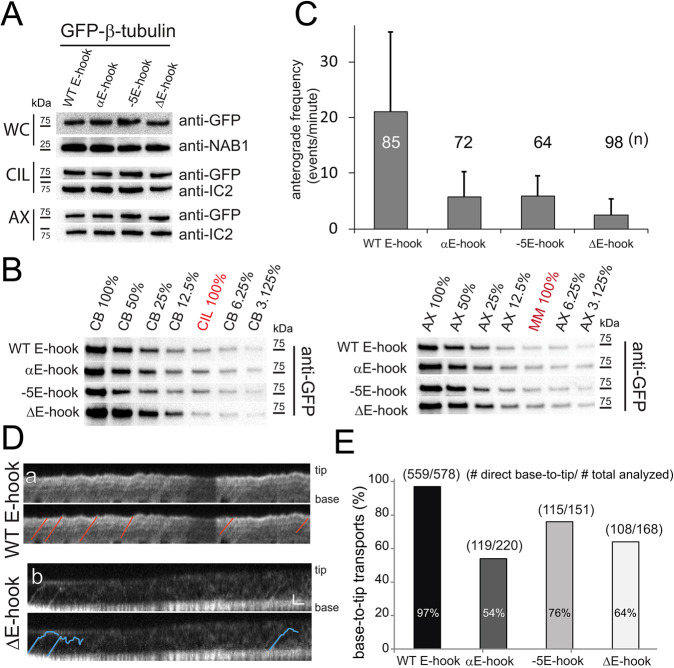
Fig. 2.
The E-hook of β-tubulin promotes transport by IFT. (A) Western blot analyses of whole cells (WC), isolated cilia (CIL), and axonemes (AX) from strains expressing GFP–β-tubulin with wild-type and modified E-hooks. Anti-GFP was used to visualize the tagged β-tubulins; antibodies to NAB1 and IC2 were used to visualize loading controls for WC and CIL/AX, respectively. (B) Left and right panels show western blots of dilution series comparing the amount of GFP–β-tubulin in the deciliated cell bodies (CB) to that in cilia (CIL), and the amount of GFP–β-tubulin in the axonemes (AX) to that in the detergent-soluble membrane and matrix fraction (MM), respectively. A value of 100% indicates that equivalents of the two fractions were loaded (i.e. one cell body per two cilia). (C) The average frequency of anterograde IFT events observed for the GFP-tagged β-tubulins. The number of regenerating cilia analyzed for each strain (n) and the s.d. are indicated. (D) Kymograms showing transport of full-length (WT E-hook) and E-hook-deficient (ΔE-hook) β-tubulin in regenerating cilia. Selected tracks are marked on the lower duplicate panel for each strain. Note the reduced frequency and reduced run length of transport events involving the truncated GFP–β-tubulin (blue lines). Scale bars: 1 s (horizontal) 1 µm (vertical). (E) The percentage of IFT events that proceeded non-stop from ciliary base to tip for wild-type (WT E-hook) and modified GFP–β-tubulins. The number of processive base-to-tip events and the total number of events analyzed are indicated.

