Fig. 4.

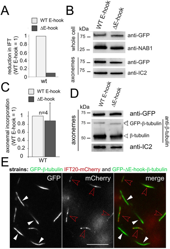
Abated IFT of E-hook-deficient tubulin does not cause a proportional reduction of its presence in the axoneme. (A) The reduction in IFT transport of truncated (ΔE-hook) versus full-length (WT E-hook) β-tubulin in control cells. The frequency observed for the full-length GFP–β-tubulin was set to 1. See Fig. 3C for the original data, s.d. and n values. (B) Western blot of control cells, comparing whole-cell and axoneme samples of the indicated strains using anti-GFP and antibodies to NAB1 and IC2, as loading controls. (C) Bar chart showing the change in the axonemal amounts of E-hook-deficient (ΔE-hook) versus full-length β-tubulin in control cells, as deduced from western blots probed with anti-GFP. The band intensity of the full-length GFP–β-tubulin was set to 1. n, number of independent cilia isolates. Data are mean+s.d. (D) Western blot comparing isolated axonemes from a wild-type strain expressing full-length or truncated β-tubulin stained with anti-GFP, anti-β-tubulin, and anti-IC2. (E) TIRF images showing the GFP and mCherry signals in cilia of live cells. Cells expressing E-hook-deficient GFP–β-tubulin (white arrowheads) and cells expressing full-length GFP–β-tubulin in the ift20-1 IFT20–mCherry background (red arrowheads) were mixed to allow for a comparison of the GFP-signal strength with the same microscope settings. Scale bar: 10 µm.
