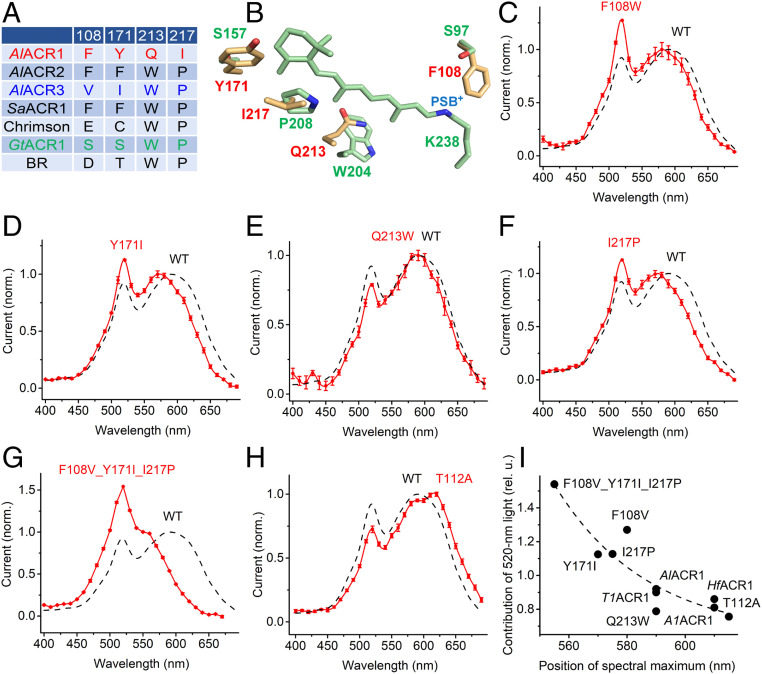
Fig. 2.
(A) The residues in the retinal-binding pocket of indicated proteins. The numbers correspond to the sequence of AlACR1. BR, bacteriorhodopsin. (B) A homology model of AlACR1 showing the side chains of the residues from A (yellow with red labels) and the chromophore and corresponding side chains of GtACR1 (green; PDB entry code: 6EDQ). PSB+, protonated Schiff base. (C–H) The action spectra of photocurrents generated by the indicated mutants of AlACR1 (red) and that of the wild type from Fig. 1B (black dashed line). (I) The dependence of contribution of the 520-nm peak (its ratio to the rhodopsin peak) on the position of the rhodopsin peak.

