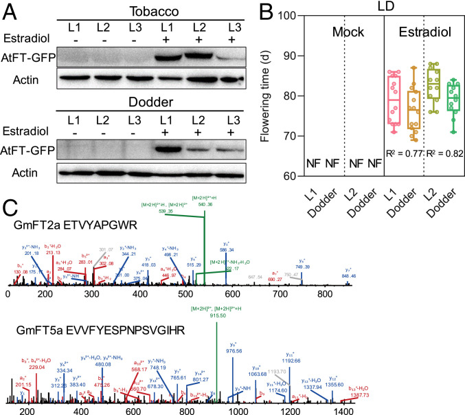
Fig. 3.
Translocation of FT proteins from hosts to C. australis. (A) Western blotting detection of AtFT-GFP in the transgenic tobacco and parasitizing dodder. Three lines (L1 to L3) of tobacco (cv Maryland Mammoth) plants, which had been transformed with AtFT-GFP driven by an estradiol-inducible promoter, were treated with estradiol or mock treated. After 48 h, tobacco leaves and stems of the parasitizing dodders were harvested for detection of AtFT-GFP protein. (B) The flowering times of the transgenic tobacco (L1 and L2) and parasitizing dodder. Tobacco plants were treated with estradiol or mock treated for 16 d (once every 2 d) (n = 21, 21, 15, 15, 14, 14, 12, and 12; from Left to Right). NF, no flowering until 120 d. L1 and L2 represent two independent lines. The horizontal bars within boxes indicate medians. The tops and bottoms of boxes indicate upper and lower quartiles, respectively. The upper and lower whiskers represent maximum and minimum, respectively. R2 indicates correlation coefficients, which were obtained from correlation analysis using the flowering times of each pair of host–dodder. (C) Mass spectrometry characterization of GmFT2a- and GmFT5a-derived peptides identified in dodder stem proteome. Dodders were grown on soybean Williams 82, which was cultivated under SD condition for about 20 d. Mass spectra indicate sequences ETVYAPGWR and EVVFYESPNPSVGIHR from GmFT2a and GmFT5a, respectively.

