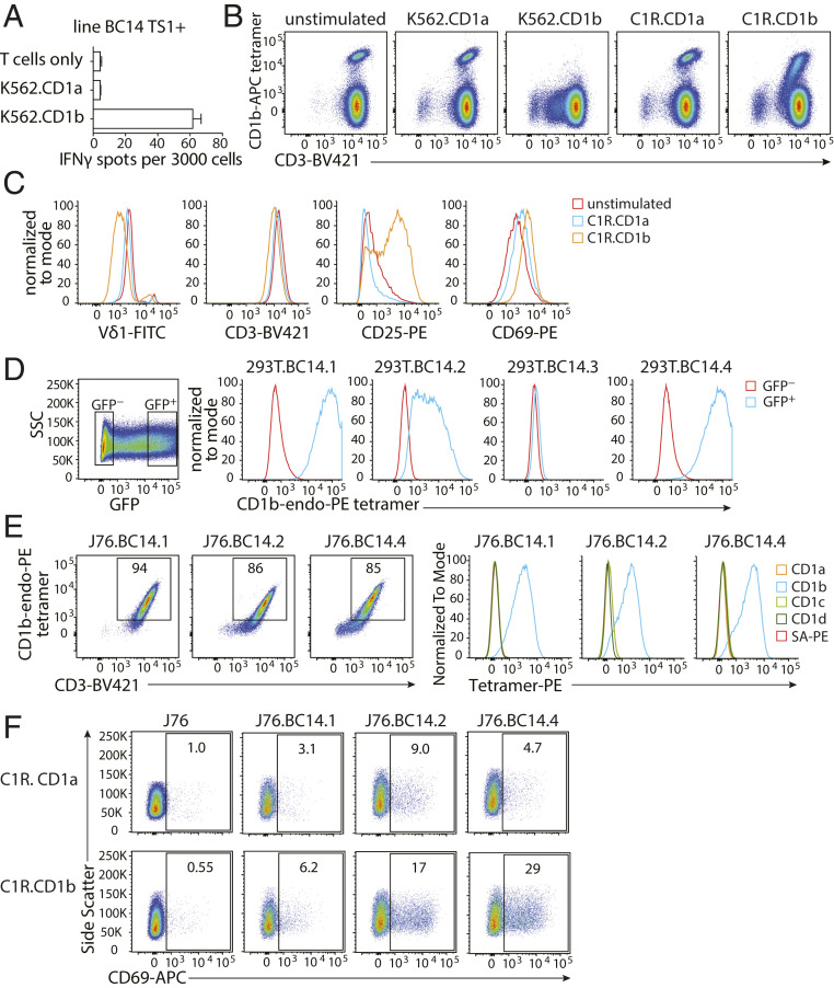
Fig. 2.
Tetramer-positive γδ T cells are autoreactive to CD1b. (A) IFN-γ ELISPOT of line TS1+ stimulated with K562.CD1a or K562.CD1b cells in the absence of exogenously added antigen. Error bars represent the SEM of triplicate wells. One representative experiment of three is shown. (B) Flow cytometry dot plots of line TS1+ after coculture with CD1a- or CD1b-expressing K562 or C1R cells. One representative experiment of three is shown. (C) Flow cytometry histograms show changes in cell surface levels of Vδ1, CD3, CD25, and CD69 on CD1b tetramer+CD3+ cells after stimulation by C1R.CD1a or C1R.CD1b cells for 20 h. (D) Flow cytometry histograms show 293T cells transfected with γδ TCRs and CD3 complex proteins stained with CD1b tetramers. The mean fluorescence intensity (MFI) of the CD1b tetramer is shown for GFP-negative and GFP-positive 293T cells. (E) Flow cytometry dot plots and histograms of J76 cells stably transduced with γδ TCRs. (F) Flow cytometry dot plots of J76 cell lines after stimulation by C1R.CD1a or C1R.CD1b cells for 20 h. One representative experiment of three experiments is shown.

