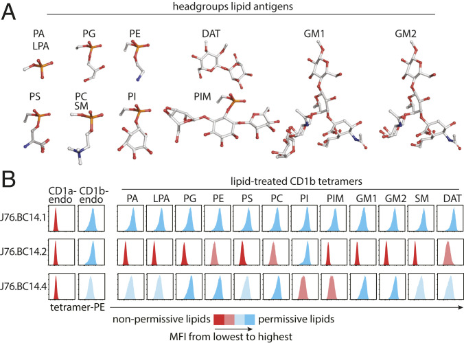
Fig. 4.
Antigen specificity of CD1b-restricted γδ TCRs. (A) Head groups of lipids used to treat CD1b monomers are shown as ball-and-stick plots. Red: oxygen; orange: phosphorus; white: carbon; blue: nitrogen. (B) Flow cytometry histograms of the indicated J76 lines stained with a panel of 13 CD1b tetramers and one CD1a tetramer carrying endogenous (endo) lipids or treated with the indicated lipid are shown. One representative experiment of two is shown.

