Fig. 4.
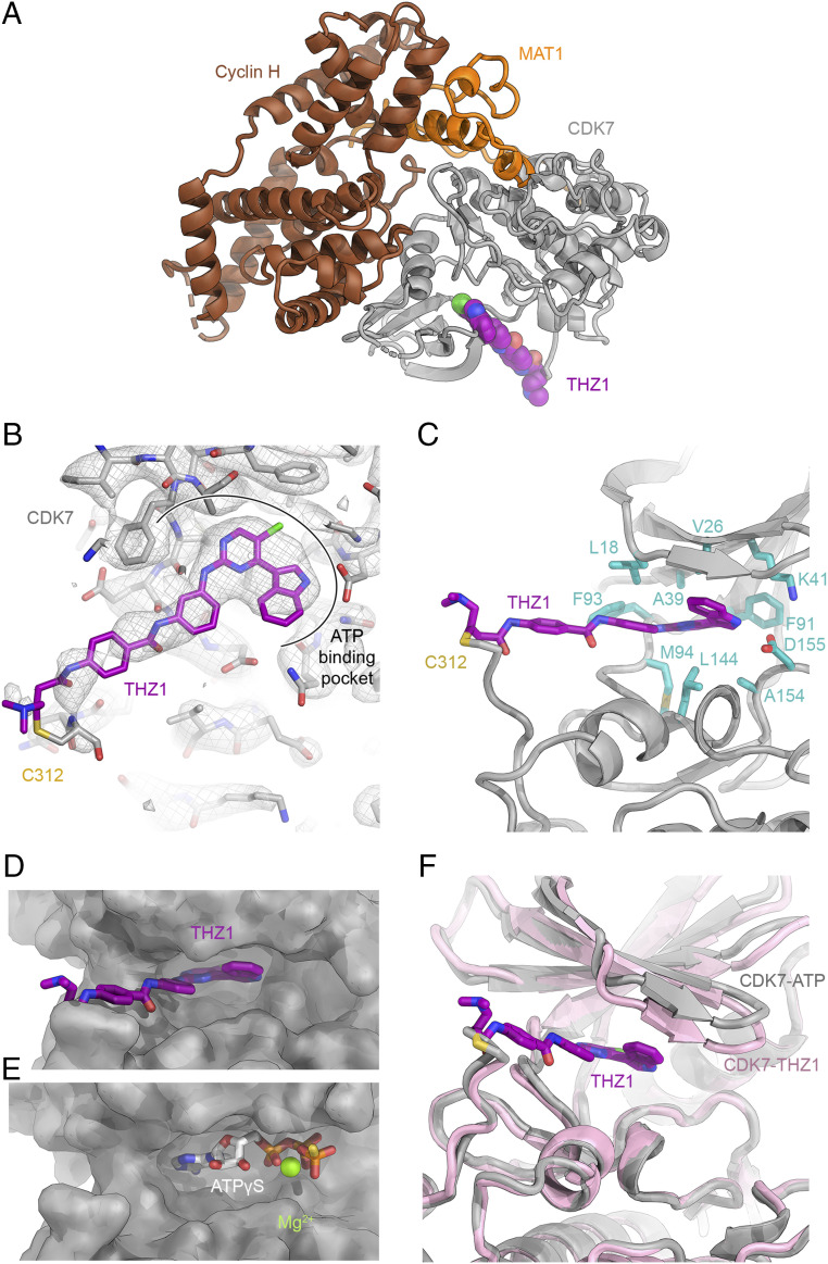
The CAK-THZ1 complex. (A) Structure of the CAK-THZ1 complex. THZ1 is shown in purple. (B) THZ1 shown in the cryo-EM density contoured at 4.5σ. THZ1 is bound in the ATP-binding pocket of CDK7 and stretches to the covalently modified cysteine C312. (C) Molecular environment of THZ1 in the nucleotide-binding pocket of CDK7, dominated by hydrophobic residues. (D) THZ1 (purple) in the nucleotide-binding cavity of CDK7. (E) ATPγS in the nucleotide-binding cavity of CDK7. The aromatic head group of THZ1 and the base and ribose of the nucleotide occupy the same site. (F) Comparison of the conformation of CDK7 in the nucleotide-bound (gray) and THZ1-bound (pink) states.

