Fig. 1.
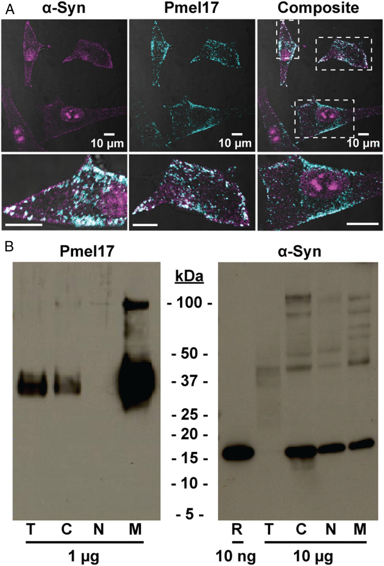
Endogenous α-syn is present in melanosomes of SK-MEL 28 human melanoma cells. (A) Confocal immunofluorescence images probed with anti−α-syn (magenta; EP1646Y) and anti-Pmel17 (cyan; HMB45) antibodies. Individual fluorescence channels were analyzed by defining a pixel threshold using the Otsu method, then using Boolean algebra to identify areas of cooccurrence (white). Dashed boxes represent areas shown below in the expanded views. (Scale bars, 10 µm.) (B) Immunoblots of total SK-MEL 28 cell lysate (T), or fractions enriched in C, N, M proteins probed with anti-Pmel17 (HMB45) or anti−α-syn (LB509) antibodies. Amount of protein loaded as indicated. Recombinant α-syn (R) is also shown for reference.

