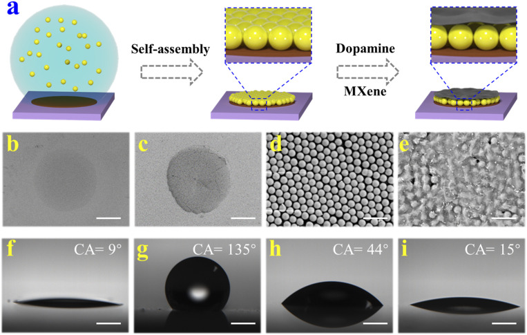
Fig. 2.
(A) Schematic illustration of the preparation process of bioinspired MXene-integrated PhC arrays. (B–E) The SEM images of (B) DA layer, (C) PhC pattern, (D) the surface of PhC pattern, and (E) the surface of MXene-integrated PhC pattern. (F–I) The water contact angles (CA) of (F) glass slide, (G) hydrophobic substrate, (H) DA layer, and (I) the MXene-integrated PhC pattern. (Scale bars are 300 μm in B and C, 1 μm in D and E, and 500 μm in F–I.)

