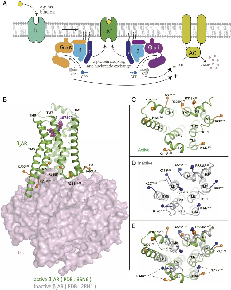
Fig. 1.
The β2AR-G protein signaling pathways. (A) The agonist-bound β2AR activates either Gs or Gi heterotrimer, which stimulates or inhibits the adenylyl cyclase activity, respectively. (B) Structure of β2AR-Gs complex (PDB ID code: 3SN6), the lysine residues that are chosen as NMR probes are shown as solid spheres, other lysine residues were mutated to arginine as described in the text. (C–E) The lysine probes undergo conformational changes during the activation of the β2AR, as shown by cytoplasmic views of active β2AR (PDB ID code: 3SN6) (C), inactive β2AR (PDB ID code: 2RH1) (D), and the overlap of active and inactive β2AR (E).

