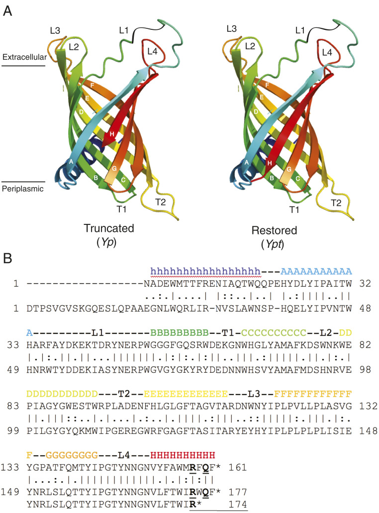
Fig. 2.
Predicted tertiary and secondary structure of PagP homologs from Y. pestis and Y. pseudotuberculosis. (A) The molecular model is shown in A without the C-terminal three amino acid residues in β-strand H (Truncated–Y. pestis) and with these residues in place (Restored–Y. pseudotuberculosis). (B) The primary structure of E. coli PagP is presented with its known secondary structural elements indicated above and with the sequence of Y. pseudotuberculosis PagP aligned below.

