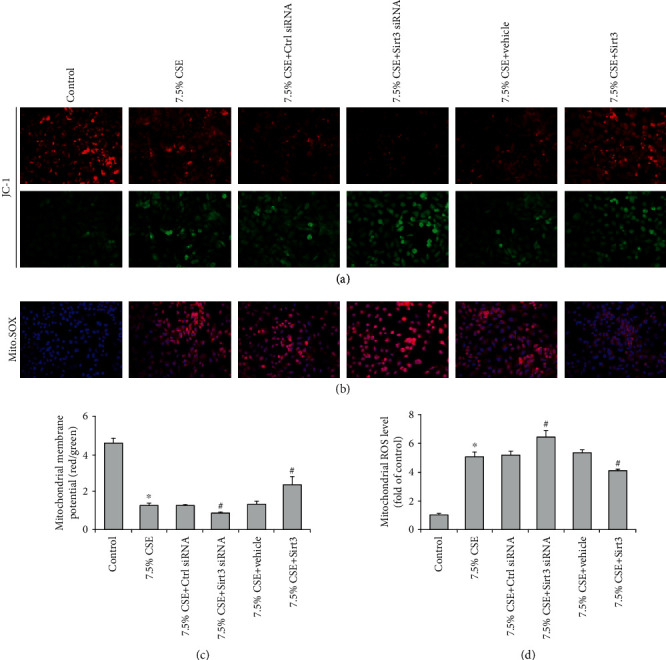
Figure 5.

Sirt3 inhibited airway epithelial mitochondrial oxidative stress induced by CSE. Airway epithelial cells were stained with JC-1 or Mito. SOX fluorescence dye and the fluorescence intensities determined by fluorescent microscope represented the levels of mitochondrial membrane potential (a) and mitochondrial ROS (b). Quantitative analysis showed CSE decreased mitochondrial membrane potential level (c) and increased mitochondrial ROS content (d), and the alterations were aggravated by Sirt3 siRNA and attenuated by Sirt3 overexpression in CSE-treated BEAS-2B cells. Results are expressed as mean ± SD from three independent experiments. ∗P < 0.01 versus control group, and #P < 0.05 versus 7.5% CSE-treated group.
