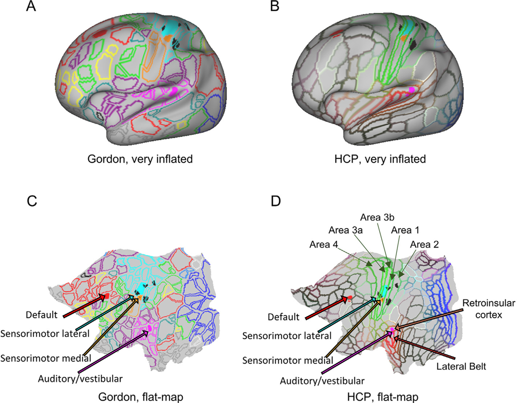
Fig. 4.
Cortical areas associated with freezing of gait. “Very inflated” and “flat map” projections of the cortical areas associated with freezing of gait on top of the delineation of regions of interest as defined by Gordon (Gordon et al., 2014) and the Human Connectome Project (HCP, (Glasser et al., 2016)). Black spots are the cortical areas in the somatosensory cortex with significant differences between PD-freezers, PD-non-freezers and controls identified by the seed analysis from the left globus pallidus (from Fig. 3(D)). Connectivity between the left globus pallidus and the areas in the sensorimotor lateral and medial cortex (Gordon ROIs 38 and 59, respectively (Gordon et al., 2014)) and between the cortical areas belonging to the Default (Gordon ROI 154) and Auditory/vestibular (Gordon 69 (Gordon et al., 2014)) networks also exhibited significant differences between groups. Legends from the HCP parcellation were included for areas overlapping the significant Gordon areas identified as relevant to FoG: Brodmann areas 1, 2, 3a, 3b, 4, 8A (dorsal), 46, lateral Belt area in the early auditory cortex and the retroinsular cortex.

