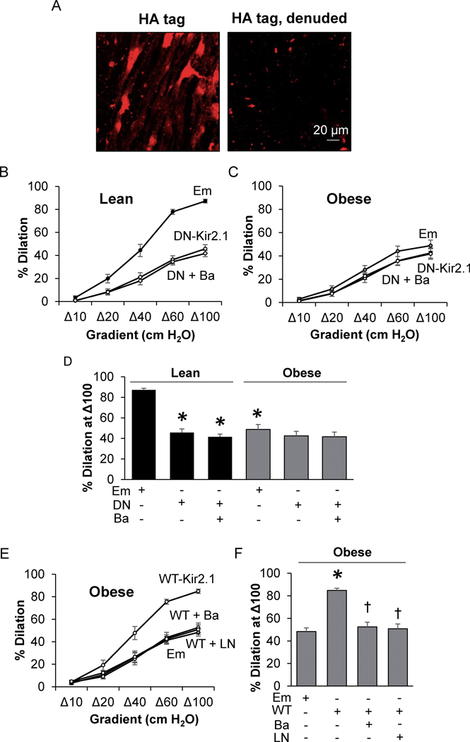
Figure 1. The loss of Kir2.1 results in FIV impairment in mesenteric adipose arteries of obese mice.
A) Images of intact and denuded en face mesenteric arteries transduced with the VE-Cadherin-WT-Kir2.1-HA tag (WT-Kir2.1)-adenovirus (AV) and stained for the HA-tag. The magnification is 25x. Ex vivo flow-induced vasodilation (FIV) measurements in arteries from B) lean and C) obese mice incubated with Empty (Em)-AV or VE-Cadherin-dominant negative-Kir2.1-AV (DN-Kir2.1) +/− BaCl2 (Ba) (n = 8). D) Analysis of FIV at a flow of Δ100; * indicates significance when compared to lean Em-AV (p<0.05). E) Ex vivo FIV measurements in arteries isolated from obese mice after incubation with either Em-AV or WT-Kir2.1-AV +/− BaCl2 or LNAME (LN) (n = 8). F) Analysis of the %dilation at Δ100; † and * indicate significance when compared to VE-Cadherin-WT-Kir2.1-HA-AV or Em-AV, respectively. Data from an equal number of male and female mice were combined in B-F.

