
Figure 1.
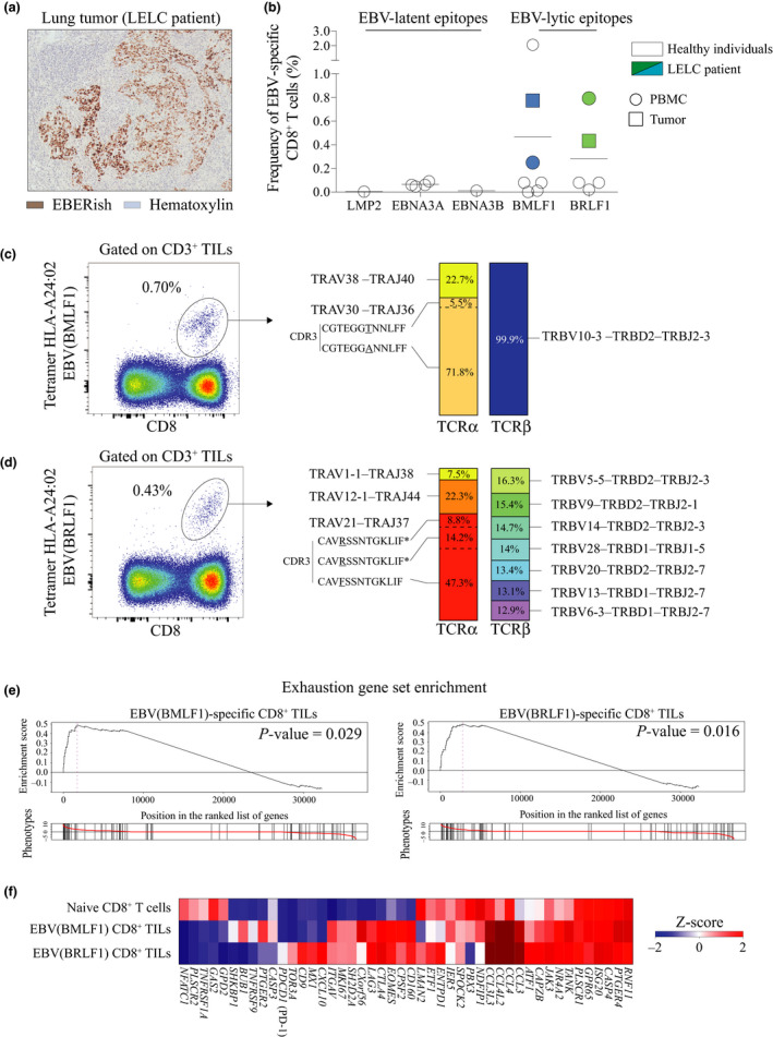
Identification of EBV‐specific CD8+ TILs in EBV‐infected LELC of the lung. (a) Immunohistochemistry staining of the lung tumor from LELC patient stained with haematoxylin (blue) and Epstein–Barr virus‐encoded small RNA in situ hybridisation (EBERish – brown) (patient A311). (b) Frequency of EBV‐specific CD8+ T cells for EBV lytic and EBV latent epitopes in HLA‐A*24: 02 LELC patient (n = 1) and healthy individuals (n = 7). (c) Flow staining of sorted EBV (BMLF1)‐specific CD8+ TILs using HLA‐A*24: 02 tetramer (DYNFVKQLF) (left panel). Frequency of tetramer‐positive cells among CD8+ TILs. TCRα and TCRβ clones’ repertoire of bulk EBV(BMLF1)‐specific CD8+ TILs (right panel). Data from LELC patient A311. (d) Flow staining of sorted EBV (BRLF1)‐specific CD8+ TILs using HLA‐A*24: 02 tetramer (TYPVLEEMF) (left panel). Frequency of tetramer‐positive cells among CD8+ TILs. TCRα and TCRβ clones’ repertoire of bulk EBV (BRLF1)‐specific CD8+ TILs (right panel), * the same CDR3 amino acid sequence but different nucleotide sequence. Data from LELC patient A311. (e) Enrichment of the gene set for exhausted T cells in naive CD8+ T cells from PBMC (flow‐sorted CCR7– CD45RO–), and EBV (BMLF1)‐ or EBV (BRLF1)‐specific CD8+ TILs. Gene position on the left indicates enrichment in EBV (BMLF1)‐ or EBV (BRLF1)‐specific CD8+ TILs. Gene position on the right indicates enrichment in naive CD8+ T cells. Data from LELC patient A311. (f) Heat map of gene set for exhaustion shown in e. Data from LELC patient A311.