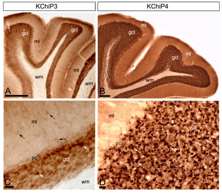
Figure 1.
Regional and cellular distribution of Kv channel interacting proteins (KChIPs) subunits KChIP3 and KChIP4 in the cerebellum. (A–D) Immunoreactivity for KChIP3 and KChIP4 in the cerebellar cortex using a pre-embedding immunoperoxidase method at the light microscopic level. Parasagittal photomicrographs of the cerebellar cortex. Immunoreactivity for both KChIP3 and KChIP4 was widely distributed in the cerebellar cortex with mostly overlapping labelling patterns. Although with some differences in intensity of labelling, strong immunoreactivity for KChIP3 and KChIP4 was found in the granule cell layer (gcl) and weaker in the molecular layer (ml) and the white matter (wm) was devoid of any staining. In the molecular layer, KChIP3 and KChIP4 was mostly neuropilar and KChIP3 labelling was also detected in cell bodies and dendrites of stellate and basket cells (black arrows). In the granule cell layer, KChIP3 and KChIP4 particularly concentrated in glomeruli (white arrows) and surrounding GCs. Scale bars: (A), 50 µm; (B), 100 µm; (C,D), 25 µm.

