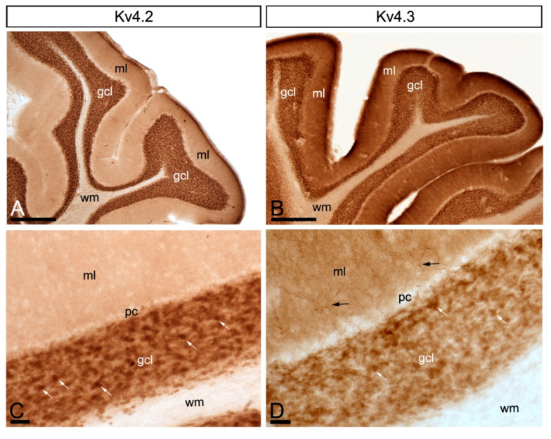
Figure 2.
Regional and cellular distribution of voltage-gated potassium (Kv) channel subunits Kv4.2 and Kv4.3 in the cerebellum. (A–D) Immunoreactivity for Kv4.2 and Kv4.3 in the rat cerebellar cortex using a pre-embedding immunoperoxidase method at the light microscopic level. Parasagittal photomicrographs of the cerebellar cortex. The strongest immunoreactivity for Kv4.2 and Kv4.3 was found in the granule cell layer (gcl). Strong immunoreactivity for Kv4.3 was also observed in the molecular layer (ml), but weaker for Kv4.2. The white matter (wm) was always devoid of any immunolabelling. Immunoreactivity for Kv4.2 and Kv4.3 in the molecular layer was mostly neuropilar, but Kv4.3 labelling was also detected in cell bodies and dendrites of basket cells (black arrows). In the granule cell layer, Kv4.2 and Kv4.3 immunolabelling particularly concentrated in glomeruli (white arrows) and surrounding GCs. Scale bars: (A,B), 50 µm; (C,D), 25 µm.

