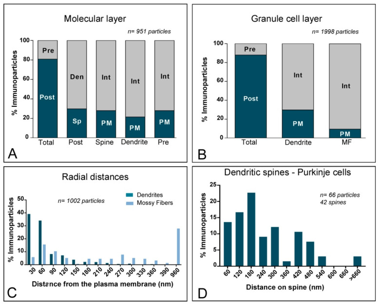
Figure 6.
Compartmentalisation of Kv channel interacting protein (KChIP) subunit KChIP4 in cerebellar cells. (A) Bar graphs showing the percentage of immunoparticles for KChIP4 at post- and pre-synaptic compartments in the molecular layer. A total of 951 immunoparticles in the molecular layer were analysed, of which 80.9% were postsynaptic and 19.1% were presynaptic. Postsynaptically, immunoparticles were detected in dendritic spines (29.8%) and in dendritic shafts (70.2%), distributed along the plasma membrane (27.9% in spines; 21.5% in dendrites) and at cytoplasmic sites (72.1% in spines; 78.5% in dendrites). (B) Bar graphs showing the percentage of KChIP4 immunoparticles at post- and pre-synaptic compartments, and along the plasma membrane and intracellular sites in dendritic shafts of granule cells and mossy fibres in the granule cell layer. A total of 1998 immunoparticles in the granular cell layer were analysed, of which 87.9% were postsynaptic and 12.1% were presynaptic. Postsynaptically, immunoparticles were detected in dendrites of GCs (87.9%), distributed along the plasma membrane (29.9%) and at cytoplasmic sites (70.1%). Presynaptically, immunoparticles were detected in mossy fibre terminals (12.1%), distributed mostly at cytoplasmic sites (90.4%) and very few along the plasma membrane (9.6%). (C) Histogram showing the radial distribution of KChIP4 immunoparticles from the plasma membrane towards cytoplasmic sites in dendrites of GCs and mossy fibres. In dendrites, immunoparticles for KChIP4 showed a skewed frequency distribution in the plasma membrane direction, but in mossy fibres, they were more equally distributed across the axoplasm. (D) Histogram showing the distribution of immunoreactive KChIP4 in relation to glutamate release sites in dendritic spines PCs. The data show the proportion of KChIP4 immunoparticles at a given distance from the edge of the postsynaptic density. About 74.2% of immunolabelled KChIP4 are located in a 60–300 nm wide band, and then the density decreased markedly further in the spine membrane.

