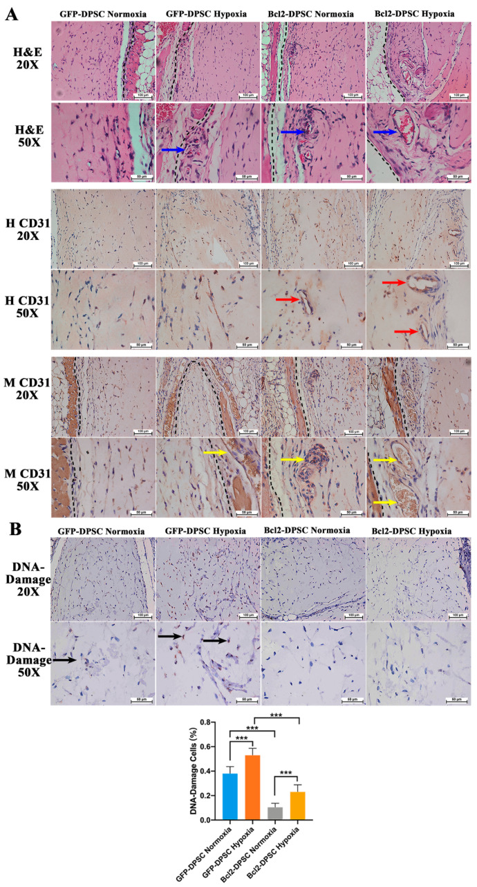
Figure 6.
In vivo Matrigel plug assay: (A) Representative microscopic images of hematoxylin and eosin (H&E) (20× and 50× magnification) and immunohistochemistry for human CD31 (H CD31; 20× and 50× magnification) and mouse CD31 (M CD31; 20× and 50× magnification) of Matrigel plugs at 7 days of implantation. Broken black lines—interface between the mouse tissue and the Matrigel plug. Blue arrows—perfused blood vessels. Red arrows—human CD31 positive non-perfused lumens. Yellow arrows—mouse CD31 positive perfused blood vessels. (B) Representative microscopic images of immunohistochemistry for DNA damage (20× and 50× magnification) and quantified percentage cells with DNA damage. Black arrows—DNA damage positive cells. *** p < 0.001 versus the corresponding controls.

