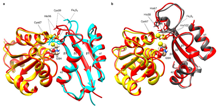
Figure 6.
Structural rearrangements in Grx5 and BolA upon dimer formation. (a) Superimposition of the backbone structure of human apo-Grx5 [PDB:2WUL] (yellow) and human BolA3 [PDB: 2NCL] (cyan) with the Fe2S2 BolA3-Grx5 complex backbone structure (red). (b) Superimposition of the backbone structure of human apo-Grx5 [PDB:2WUL] (yellow) and human BolA1 [PDB:5LCI] (dark gray) with the Fe2S2 BolA1-Grx5 complex backbone structure (red). The GSH molecule and the Fe2S2 cluster are represented as balls and sticks. The invariant C-terminal His (His 96 in BolA3 and His 102 in BolA1), His 67 in BolA1, Cys 59 in BolA3, and Cys 67 in human Grx5 residues are shown.

