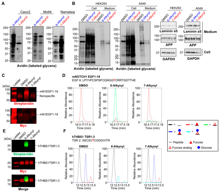
Figure 2.
Differential glycoprotein labeling with 6-Alk-Fuc and 7-Alk-Fuc. (A) Caco-2, Molt4, and Namalwa cells were treated with peracetylated 6-Alk-Fuc, 7-Alk-Fuc, or DMSO. Incorporated Fuc analogs in the cell lysates were biotinylated by click chemistry, and the labeled glycans on proteins were detected by blotting with HRP-streptavidin. (B) HEK293 and A549 cells were treated with peracetylated 6-Alk-Fuc, 7-Alk-Fuc, or DMSO. Left 2 panels: Incorporated Fuc analogs in the cell lysates or secreted proteins were biotinylated by click chemistry, and the labeled glycans on proteins were detected by blotting with HRP-streptavidin. Right 2 columns: Proteins in the cell lysates and secreted into the culture media were western blotted with anti-laminin alpha 5, anti-APP, or anti-GAPDH. (C) HEK293T cells were transfected with mouse NOTCH 1 (mN1) EGF1-18 plasmid and incubated with 50 μM peracetylated 6-Alk-Fuc or 7-Alk-Fuc for 4 days. mNOTCH1 EGF1-18 was purified from media and subjected to CuAAC with azido-biotin probe to examine Fuc analog incorporation. The samples were analyzed by Western blot. Top panel: Probed with streptavidin; bottom panel, probed with anti-Myc. (D) mN1 EGF1-18 was prepared as in C, digested with V8 protease, and the resulting peptides analyzed by mass spectrometry. An extracted ion chromatogram (EIC) of glycoforms of a peptide from mNOTCH1 EGF6 was prepared. Spectra for these ions are in Supplementary Figure S1. Black line, unmodified; red line, Fuc modified; green line, 6-Alk-Fuc or 7-Alk-Fuc modified. (E) HEK293T cells were transfected with hTHBS1 TSR1-3 plasmid and incubated with 50 μM peracetylated 6-Alk-Fuc or 7-Alk-Fuc for 3 days. hTHBS1 TSR1-3 was purified from secreted media and subjected to CuAAC with azido-biotin probe to examine Fuc analog incorporation. The samples were analyzed by Western blot. Top panel: Probed with streptavidin; middle panel, probed with anti-Myc; bottom panel, merged. (F) hTHBS1 TSR1-3 was prepared as in E, digested with trypsin and chymotrypsin, and the resulting peptides analyzed by mass spectrometry. An EIC of the different glycoforms of a peptide from hTHBS1 TSR2 was prepared. Spectra for these ions are in Supplementary Figure S2. Black line, unmodified; red line, Fuc modified; blue line, glucose (Glc)-Fuc modified; green line, 6-Alk-Fuc or 7-Alk-Fuc modified; aqua line, Glc-6-Alk-Fuc or Glc-7-Alk-Fuc.

