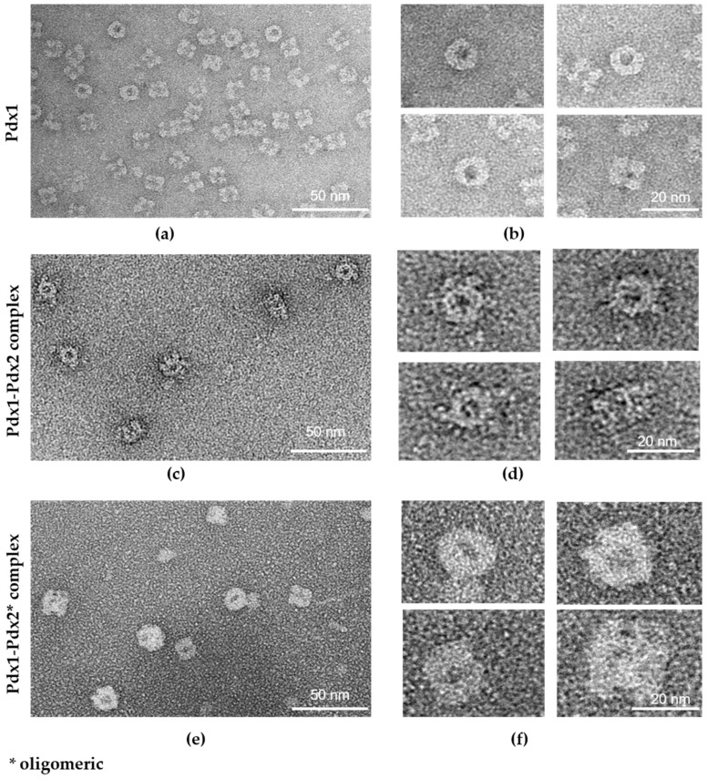
Figure 5.
Transmission electron micrographs of negatively stained P. vivax pyridoxal phosphate (PLP) synthase proteins. Images on the right in (b,d,f), alongside a 20 nm scale bar, show a zoom-in of a representative class of averaged sample pictures shown on the left in (a,c,e). (a,b) Dodecameric Pdx1, top view, and side view with random orientations of the particles; (c,d) dodecameric Pdx1 in complex with Pdx2; for complex formation, a monomeric Pdx2 solution was provided. Pdx complex particles generally only with partially bound Pdx2 were observed and saturated (12:12) Pdx complex was rarely observed; (e,f) dodecameric Pdx1 in complex with Pdx2 oligomers. The Pdx complex was predominantly saturated with Pdx2, showing a slightly larger dimension than those observed for the Pdx complexes shown in (c,d), demonstrating the larger hydrodynamic radius of the corresponding Pdx complex observed by DLS (Figure 2b). * oligomeric Pdx2.

