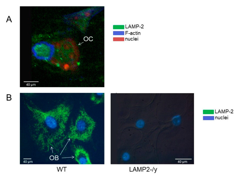
Figure 1.
Lysosome associated membrane protein 2 (LAMP-2) localization in osteoclast and osteoblasts. LAMP-2 (visualized with alexa-488 (green), localization in (A) Confocal image of a wild type osteoclast (OC) on a cortical bone slice. The green LAMP-2 label is mainly present in the area surrounded by actin (visualized with phalloidin-alexa-633, in blue). This area is considered as the ruffled border area. Nuclei were stained with propidium iodide (red). (B) Wild-type (left) and LAMP-2-/y (right) osteoblasts (OB) cultured on plastic. Images made by a converted fluorescence microscope. The green LAMP-2 label is present throughout the cytoplasm of wild type cells, but completely absent in the LAMP-2-/y cells. Nuclei were stained with di-amidino-2-phenylindole-dihydrochloride (DAPI).

