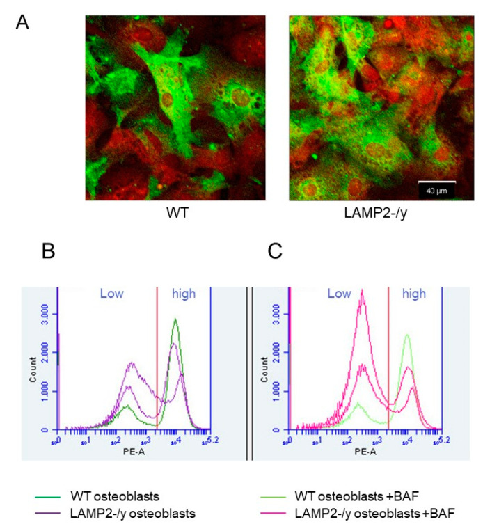
Figure 6.
Immunolocalization and Fluorescence-activated cell sorting (FACS) analysis of RANKL in wild-type and LAMP-2 -/y osteoblasts. (A) WT and LAMP-2-/y osteoblasts were labeled for RANKL and visualized with alexa-488 (green), the nuclei were stained with propidium iodide (red). About 50% of the osteoblasts from both, WT and LAMP-2 -/y mice are intensely labeled for RANKL. (B) FACS analysis showed that membrane-bound RANKL (labeled with Phycoerythrin (PE)) was not highly expressed by all osteoblasts. Part of the osteoblasts show low membrane labelling (left side of the graph (Low)). WT osteoblasts (purple line) have more RANKL on their plasma membrane than LAMP-2-/y osteoblasts (dark blue line in graph B right side (high)). (C) When osteoblasts were incubated with bafilomycin, a lower number of cells were found in the high membrane labeling fraction. This is seen at the right side of the figure (high). This counts for WT as well as LAMP-2-/y cells. WT with bafilomycin (pink line), LAMP-2-/y with bafilomycin (light green line).

