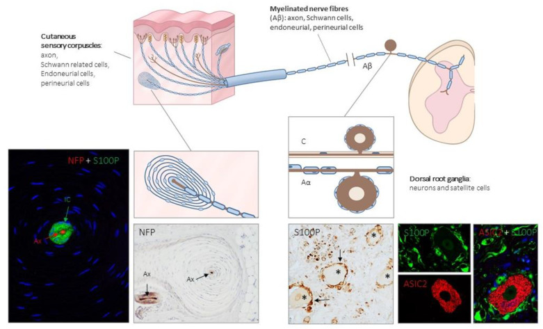
Figure 1.
Schematic representation of the afferent innervation of mammalian glabrous skin. Glabrous skin is supplied by myelinated and non-myelinated nerve fibers (Aβ, Aδ, C), originated from large, intermediate and small sized neurons (low-threshold mechanoreceptors (LTMRs) and nociceptors) localized in the dorsal root ganglia (DRG). Aβ nerve fibers end in the dermis forming different morphotypes of sensory corpuscles. Photos on the left side correspond to sections of Pacini’s corpuscles immunostained for neurofilament proteins (NFP) and S100 protein (S100P) to, respectively, label the axon (Ax; red immunofluorescence) and the Schwann-related cells (IC: inner core; green fluorescence). Right side photos correspond to a section of human lumbar DRG—immunostained for S100P and acid-sensing ion channel protein 2 (ASIC2). S100 protein labels satellite cells (arrows and green fluorescence) while neuronal cell bodies (asterisks and red fluorescence) display ASIC2 positivity.

