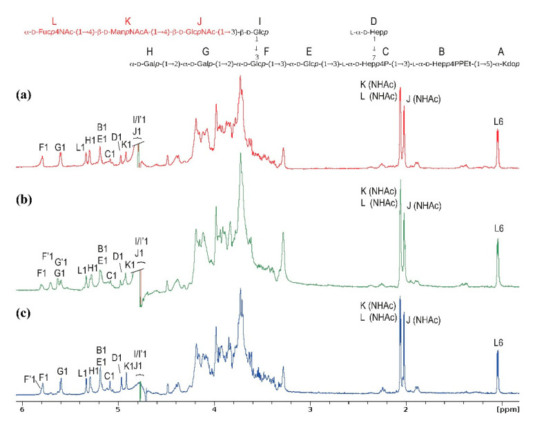
Figure 3.
The 600 MHz 1H Nuclear Magnetic Resonance (NMR) spectra of the [ECA]-core OS glycoforms identified in fractions 5 of (a) E. coli R1; (b) E. coli PCM 209 (O39); (c) S. sonnei phase II LOS preparations [7,8]. The ECA repeating unit is colored in red. The capital letters refer to carbohydrate residues of the [ECA]-core OS, as shown in the inset structure. Letters with a prime sign denote residues of trace amounts of the core OS devoid of ECA. Chemical shift assignment for E. coli R1 is present in Table 2.

