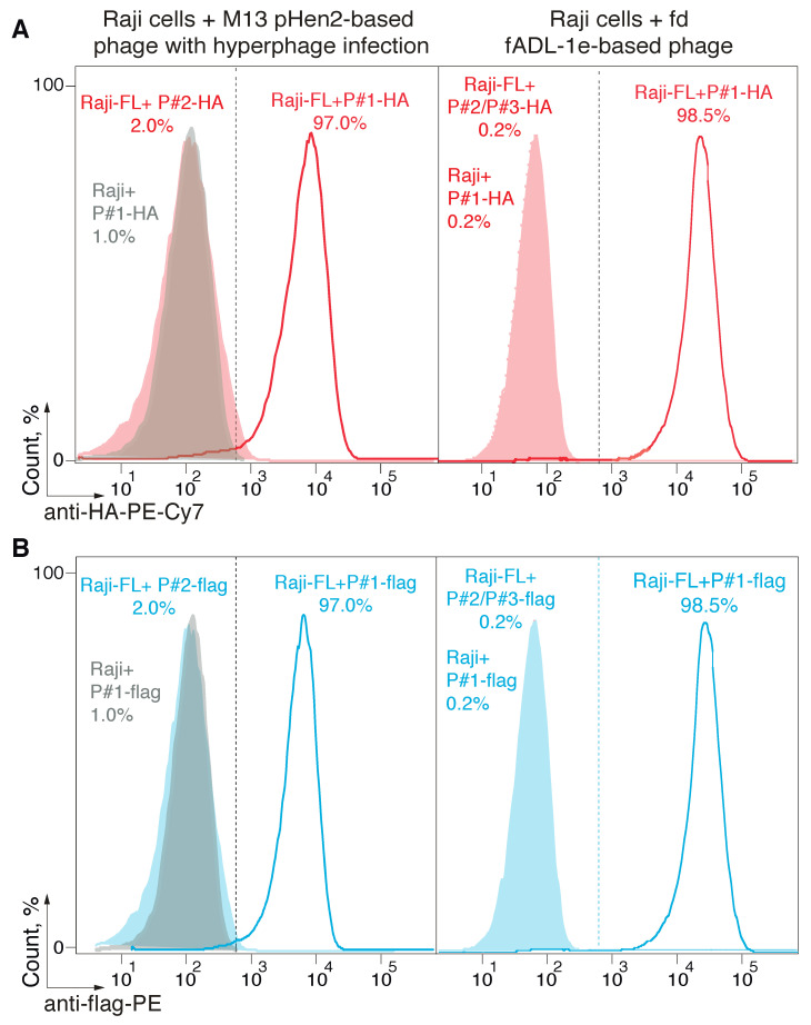
Figure 2.
Detection of the receptor-ligand interaction with the fADL-1e-based and pHen2-produced bacteriophages. Ligand–receptor interaction was studied by the flow cytometry using Raji cells expressing a membrane-tethered BCR in a single-chain format (Raji-FL cells), and filamentous bacteriophages carrying its peptide ligand (P#1). Non-transduced Raji and Raji-FL cells were incubated with filamentous phages exposing P#1-peptide fused with HA-tag (A) or 3xFLAG (B) at a concentration of 1 × 10¹² or 5 × 10¹² phage particles per mL for fADL-1e-based (right) or pHen2-based (left) protocols, respectively. Phages exposing irrelevant P#2 and P#3 peptides were used as a negative control. Fluorescence signals are plotted on the x-axis, and the percentage of the recorded events is on the y-axis. Histograms for pHen2-based + hyperphage system (left) show the cut-off gate at a false-positive signal level of 2%. Histograms for fADL-1e-based (right) show the cut-off gate at a false-positive signal level of 0.2%.

