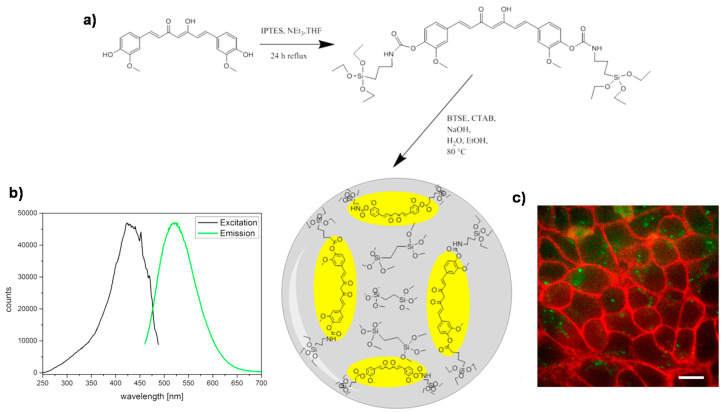
Figure 15.
(a) Preparation of PMOs (yellow-colored represents the curcumin-based units) without using additional silica; (b) Fluorescence excitation and emission spectra of the nanomaterial in PBS buffer; (c) Cellular uptake of these PMOs (green) after 24 h on HeLa cells (red: Wheat Germ Agglutinin, WGA 647 membrane staining); scale bar represents 10 μm. Adapted from [56] (https://doi.org/10.1016/j.micromeso.2015.12.006), with permission from Elsevier.

