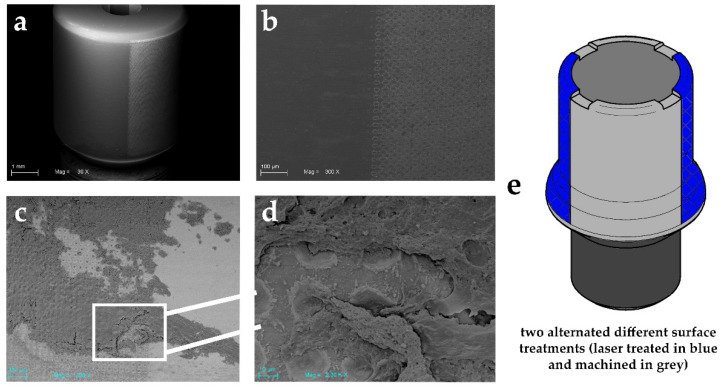
Figure 1.
SEM images showing an experimental healing abutment used in this study: (a) showing the two different alternated surface treatments (laser treated on right and machined on left). Scale bar: 1 mm; (b) high magnification of these two surfaces topography. Scale bar: 100 μm; (c) soft tissue adhesion on both the studied surfaces. Please note the high quantity on the laser-treated surface compared to the machined one Scale bar: 100 μm; (d) higher magnification of the selected zone (Scale bar: 10 μm) and its interaction with soft tissue after biopsy; (e) technical design of experimental healing abutment.

