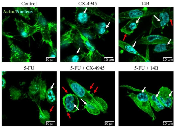
Figure 5.
Confocal laser scanning microscopy examinations of MDA-MB-231 human breast cancer cells, untreated (control) or treated with 15 µM 5-FU, 2 µM 14B, and 5 µM CX-4945 used separately or in combinations for up to 72 h, then fixed and stained with Alexa Fluor 488 phalloidin (F-actin, green fluorescence) and Hoechst 342 (nuclei, cyan fluorescence). White arrows, abnormal cell nuclei (irregular nuclei with condensed chromatin); red arrows, blebs.

