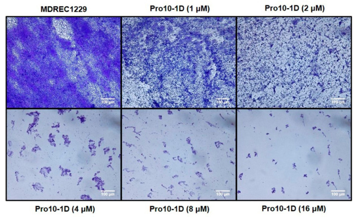
Figure 8.
Light microscopic images representing the antibiofilm effect of Pro10-1D against MDREC 1229: MDREC 1229 cells were allowed to adhere the polystyrene surface of the culture plate and were then exposed to Pro10-1D (0–16 μM) for 16 h. Strains were cultured in Mueller–Hinton (MH) media, and biofilm formation was evaluated by crystal violet staining. Pro10-1D-exposed biofilms appeared thinner and scantier than untreated MDREC 1229; these effects were dose-dependent. Scale bar, 100 μm.

