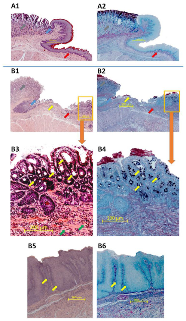
Figure 7.
Microscopic appearance of esophageal mucosa, gastroesophageal junction (GEJ) and gastric cardia stained with haematoxylin/eosin (H&E) or alcian blue/periodic acid-Schiff (AB/PAS) in representative rats without (A1,A2) or with an esophagogastroduodenal anastomosis (B1–B6). Grey arrow points out gastric mucosa, blue arrow points out GEJ, red arrow indicates esophageal mucosa, yellow arrow indicates esophageal ulceration, orange frame shows Barrett’s-like lesions (A1,A2,B1,B2). (B3,B4) show high resolution images of Barrett’s-like lesions where yellow arrows indicate goblet cells and green arrows indicate fibrosis. Yellow arrows indicate epithelial hyperplasia (B5,B6).

