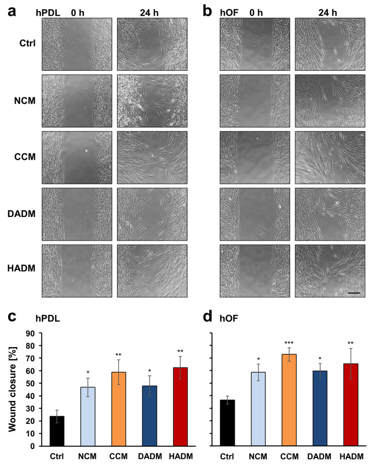
Figure 2.
Enhanced wound healing potential of primary human oral cell types covered with four different collagen matrices. (a,b) Migration of hPDL (a) and hOF (b) cells toward an wound gap with a 500 µm width generated by seeding cells in a 2-well ibidi culture inserts, in the absence (Ctrl) or in the presence of NCM, CCM, DADM, and HADM matrices placed over the cells. Representative images of the two cell types in each of the experimental groups at 0 and 24 h are shown. Scale bar, 500 μm. (c,d) Bar charts presenting quantification of the wound healing potential of hPDL (c) and hOF (d) cells, in the absence or presence of collagen matrices, by measuring the cell-free area at 0 and 24 h. Percentage of wound closure was calculated as described in the Materials and Methods section. Data represent means ± SD from three independent experiments performed with three different cell donors, in triplicates. Significant differences to the control cells, *** p < 0.001, ** p < 0.01, * p < 0.05.

