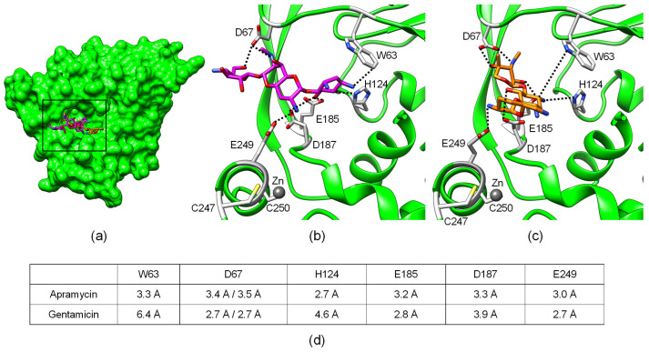
Figure 2.
Crystal structure of AAC(3)-IV in complex with apramycin (PDB ID: 6MN4) or gentamicin (PDB ID: 6MN3). (a) Structural overview of full protein showing superimposition of bound substrates apramycin (magenta) and gentamicin (orange). Detailed view of (b) apramycin and (c) gentamicin in the binding pocket. Amino acids modified in this study are highlighted in gray. Residues D67, D187, and E249 are differently rotated in the apramycin versus the gentamicin structure. (d) Shortest intermolecular distances between amino acid side chains and ligand, corresponding to dashed lines in (b,c).

