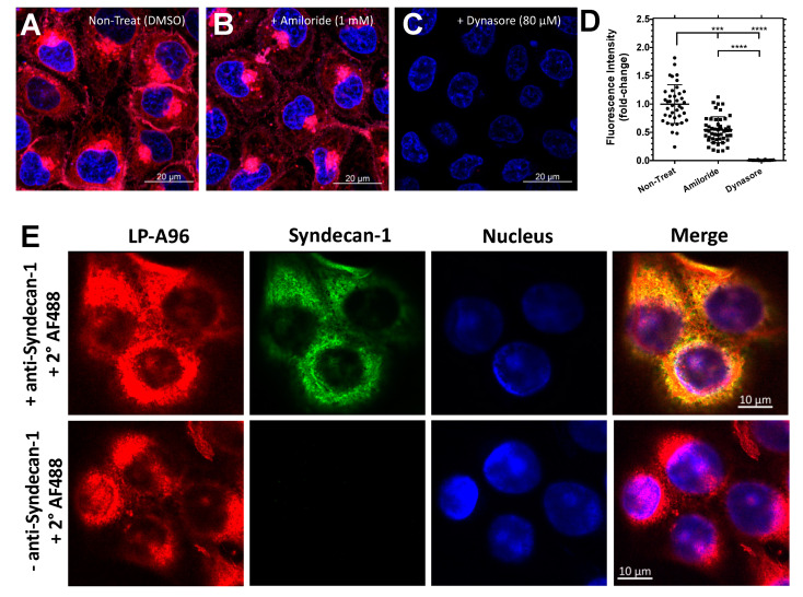
Figure 6.
LP-A96 is internalized via dynamin-dependent endocytosis and colocalized with syndecan-1 in corneal epithelial cells. (A–D) HCE-T cells were treated with either DMSO (vehicle control), amiloride (1 mM), or dynasore (80 μM) prior to addition of 1 μM rhodamine-labeled LP-A96. (D) Quantified fluorescence intensity per cell under each treatment. n = 39 for DMSO, n = 49 for amiloride, n = 48 for dynasore. Representative images are shown from the three independent experiments. (E) LP-A96 was colocalized with syndecan-1 in HCE-T cells. (Pearson’s correlation = 0.8, n = 32). 1 μM rhodamine-labeled LP-A96 were incubated with the cells for 15 min at 37 °C prior to fixation. Secondary immunofluorescence revealed apparent colocalization of rhodamine-labeled LP-A96 and Syndecan-1. Anti-syndecan-1 antibody was further detected by Alexa Fluor 488 conjugated secondary antibody. Nuclei were stained with DAPI (4′,6-diamidino-2-phenylindole). A one-way ANOVA followed by multiple comparisons was used for statistical comparison. **** p < 0.0001, *** p < 0.001. Mean ± SD.

